Publications
Light Sheet-Based Laser Patterning Bioprinting Produces Long-Term Viable Full-Thickness Skin Constructs
Tissue engineering holds great promise for biomedical research and healthcare, offering alternatives to animal models and enabling tissue regeneration and organ transplantation. 3D bioprinting stands out for its design flexibility and reproducibility. Here, an integrated fluorescent light sheet bioprinting and imaging system is presented that combines high printing speed (0.66 mm3/s) and resolution (9 µm) with light sheet-based imaging. This approach employs direct laser patterning and a static light sheet for confined voxel crosslinking in photocrosslinkable materials. The developed bioprinter enables real-time monitoring of hydrogel crosslinking using fluorescent recovery after photobleaching (FRAP) and brightfield imaging as well as in situ light sheet imaging of cells. Human fibroblasts encapsulated in a thiol-ene click chemistry-based hydrogel exhibited high viability (83% ± 4.34%) and functionality. Furthermore, full-thickness skin constructs displayed characteristics of both epidermal and dermal layers and remained viable for 41 days. The integrated approach demonstrates the capabilities of light sheet bioprinting, offering high speed, resolution, and real-time characterization. Future enhancements involving solid-state laser scanning devices such as acousto-optic deflectors and modulators will further enhance resolution and speed, opening new opportunities in light-based bioprinting and advancing tissue engineering.
Upgrading a Consumer Stereolithographic 3D Printer to Produce a Physiologically Relevant Model with Human Liver Cancer Organoids
A widespread application of 3D bioprinting in basic and translational research requires accessibility to affordable printers able to produce physiologically relevant tissue models. To facilitate the use of bioprinting as a standard technique in biology, an open-source device based on a consumer-grade 3D stereolithography apparatus (SLA) printer is developed. This SLA bioprinter can produce complex constructs that preserve cell viability and recapitulate the physiology of tissues. The detailed documentation of the modifications apported to the printer as well as a throughout performance analysis allow for a straightforward adoption of the device in other labs and its customization for specific applications. Given the low cost, several modified bioprinters could be simultaneously operated for a parallelized tissue production. To showcase the capability of the bioprinter, constructs consisting of patient-derived cholangiocarcinoma organoids encapsulated in a gelatin methacrylate (GelMA)/polyethylene glycol diacrylate (PEGDA) hydrogel are produced. A thorough characterization of different GelMA/PEGDA ratios reveals that the mechanical properties of the bioprinted tumor model can be accurately fine-tuned to mimic a specific tumor micro-environment. Immunofluorescence and gene expression analyses of tumor markers confirm that the bioprinted synthetic hydrogel provides a flexible and adequate replacement of animal-derived reconstituted extracellular matrix.
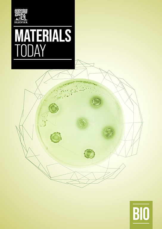
Gravitational forces and matrix stiffness modulate the invasiveness of breast cancer cells in bioprinted spheroids
.webp)
Combined Imaging and Soft and Complex 3D Bioprinting Using Light Sheet Fluorescence Microscopy
Engineered tissues are large structures made up of different cell types within a three-dimensional scaffold mimicking the extracellular matrix. Over days and weeks in culture, the cells establish tissue-like cell-cell and cell-matrix contacts forming complex internal structures with an inhomogeneous refractive index. Because of the refractive index mismatch, bioengineered tissues strongly scatter light as their native counterparts and are optically opaque at most wavelengths used in light microscopy. Moreover, large bioengineered samples are challenging to image with fluorescence microscopy due to their size and the time required to record their full volume. This also relates to the fact that most microscopes waste the sample’s available photon budget—the quantity of photons that the sample can emit before deteriorating—by exposing it to high-intensity light that induces photodamage and photobleaching. Fluorescence imaging is particularly affected by photon wasting. Among the available fluorescence microscopies, light sheet fluorescence microscopy (LSFM) is optimally suited to image large three-dimensional specimens. LSFM drastically reduces photodamage by placing the excitation and detection optics orthogonally and not collinearly, at variance with confocal microscopy. Besides volumetric fluorescence imaging, also light-based three-dimensional (3D) bioprinting techniques can greatly benefit from light sheet illumination as the improved photon budget and speed of light sheet microscopy can be leveraged in these bioprinting processes. The lower light dose combined with fast patterning ensure higher cell viability in the final constructs. A further benefit of the light sheet bioprinter is that three-dimensional imaging of the constructs can be performed on the same device. In fact, the cells need to be imaged both during printing and post-printing to monitor their collective self-organization in the bioengineered tissue. Using fluorescent probes, assessment of bioprinted samples can be performed following a straightforward workflow. This chapter present a short summary on the application of light sheet technology for imaging and bioprinting of bioengineered samples. It also presents an overview of its application in the bioengineering field, including work on soft 3D bioprinting and 3D patterning of tissue constructs.